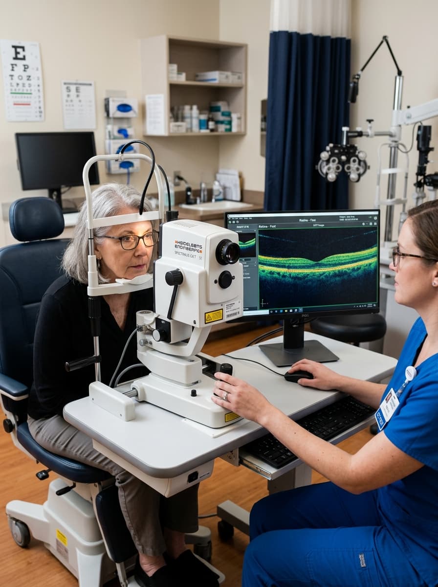
OCT (Optical Coherence Tomography - Spectral Domain)
Sub-Micron Cross-Sectional Retinal & Optic Nerve Tomography
Live Surgical Media & Consultant Demonstrations
Observe senior fellowship eye surgeons performing OCT (Optical Coherence Tomography - Spectral Domain) with gold-standard international platforms.
Doctor Surgical Playlist (0 items)
Have your eye evaluated directly by the specialist performing this procedure at our Shah Jamal clinic.
Comprehensive Clinical Overview
The optical equivalent of an in-vivo microscopic retinal biopsy. Infrared light waves generate cross-sectional 3D images of all 10 retinal layers down to 3-5 microns, revealing microscopic fluid accumulations before vision drops.
Patients with diabetic macular edema, age-related macular degeneration (AMD), macular holes, central serous chorioretinopathy, or glaucoma suspects.
Why Choose The Eyes for this Procedure:
Common Symptoms & Clinical Indications
Patients suffering from these visual or physical complaints are typically recommended for this intervention:
Heidelberg Spectralis / Topcon 3D OCT Spectral Domain Scanner
85,000 A-scans/second with confocal scanning laser ophthalmoscopy
Step-by-Step Surgical & Diagnostic Protocol
01. Automated 3D Macular Volume Scan
High-speed infrared beam scans central macula without bright blinding flashes.
02. Retinal Layer Segmentation
Computerized segmentation measures exact retinal nerve fiber layer (RNFL) and foveal thickness.
03. Progression Comparison Tracking
Compares against previous clinic visits to evaluate response to anti-VEGF injections or glaucoma drops.
Ready to Consult for OCT (Optical Coherence Tomography - Spectral Domain)?
Consultations are held at 162-A Shah Jamal Lahore. We allocate conflict-free 20-minute consultation slots with transparent, honest fees and zero corporate markups.