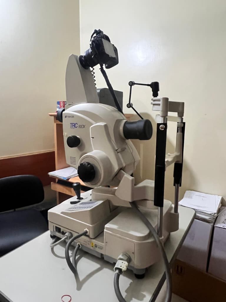
High-Resolution Digital Fundus Camera
True-Color 50-Megapixel Retinal & Optic Disc Documentation
Live Surgical Media & Consultant Demonstrations
Observe senior fellowship eye surgeons performing High-Resolution Digital Fundus Camera with gold-standard international platforms.
Doctor Surgical Playlist (0 items)
Have your eye evaluated directly by the specialist performing this procedure at our Shah Jamal clinic.
Comprehensive Clinical Overview
Captures true-color high-resolution digital photographs of the optic nerve head, macula, and peripheral retinal vasculature. Enables side-by-side progression tracking across years to detect microscopic changes early.
All diabetic, hypertensive, and glaucoma patients requiring high-resolution permanent photographic baselines to detect subtle yearly changes.
Why Choose The Eyes for this Procedure:
Common Symptoms & Clinical Indications
Patients suffering from these visual or physical complaints are typically recommended for this intervention:
Topcon Digital Retinal Camera with Tele-Ophthalmology Archiving
Multi-spectral RGB flash sensor, non-mydriatic & mydriatic widefield optics
Step-by-Step Surgical & Diagnostic Protocol
01. Automated Eye Alignment
Infrared sensor aligns with pupil without blinding flash during initial positioning.
02. Flash Image Capture
A brief low-intensity flash records high-definition true-color 50-megapixel retinal topography.
03. Side-by-Side Year-over-Year Comparison
Digital clinical archive compares previous year images to verify zero disease progression.
Ready to Consult for High-Resolution Digital Fundus Camera?
Consultations are held at 162-A Shah Jamal Lahore. We allocate conflict-free 20-minute consultation slots with transparent, honest fees and zero corporate markups.