FFA (Fundus Fluorescein Angiography)
Dynamic Microvascular Dye Angiography for Retinal Vascular Disease
Live Surgical Media & Consultant Demonstrations
Observe senior fellowship eye surgeons performing FFA (Fundus Fluorescein Angiography) with gold-standard international platforms.
Doctor Surgical Playlist (0 items)
Have your eye evaluated directly by the specialist performing this procedure at our Shah Jamal clinic.
Comprehensive Clinical Overview
A safe vegetable-based fluorescent dye is injected into an arm vein. Rapid high-resolution retinal photos capture dye circulating through retinal and choroidal vessels, highlighting microaneurysms, ischemia, and leakages.
Diabetic patients, hypertension patients, and individuals with suspected choroidal neovascularization requiring laser or anti-VEGF injection planning.
Why Choose The Eyes for this Procedure:
Common Symptoms & Clinical Indications
Patients suffering from these visual or physical complaints are typically recommended for this intervention:
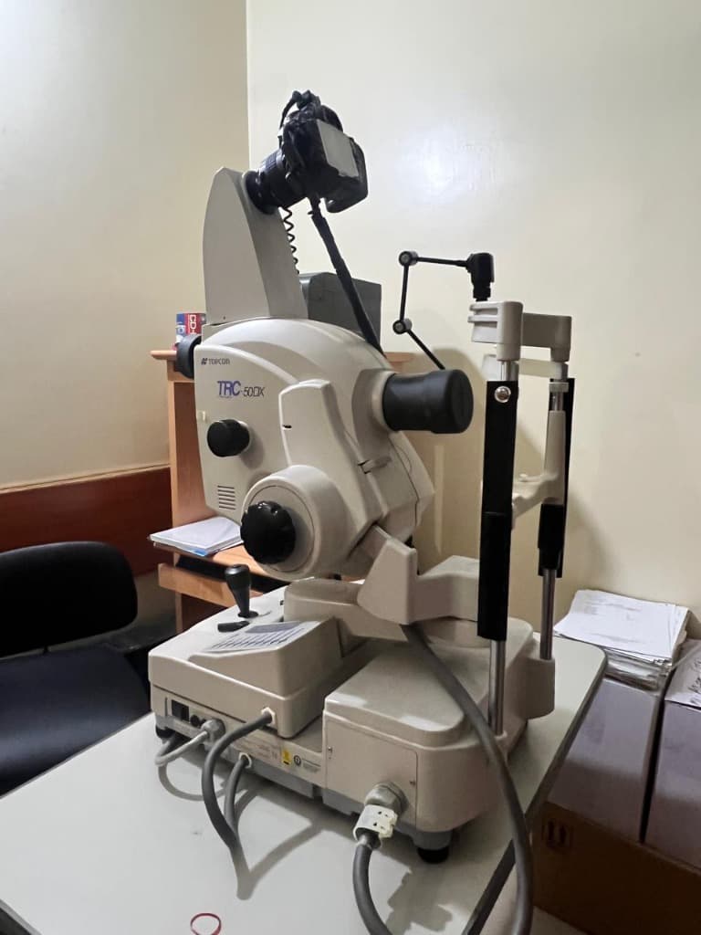
Topcon TRC-50DX High-Resolution Digital Retinal Angiography System
Dual excitation filters, rapid digital sensor with high-speed serial angiography
Step-by-Step Surgical & Diagnostic Protocol
01. Pupil Dilation & Baseline Fundus Photo
Pupils are dilated and high-resolution color baseline photographs of the retina are taken.
02. Fluorescein Dye Administration
Sodium fluorescein dye is administered intravenously via arm cannula by trained medical staff.
03. Serial Multi-Phase Digital Imaging
Early arterial, capillary, and late transit phases capture micro-vascular leaks and non-perfusion zones.
Ready to Consult for FFA (Fundus Fluorescein Angiography)?
Consultations are held at 162-A Shah Jamal Lahore. We allocate conflict-free 20-minute consultation slots with transparent, honest fees and zero corporate markups.