B-Scan Ophthalmic Ultrasound
High-Frequency Acoustic Imaging for Dense Media Opacities
Live Surgical Media & Consultant Demonstrations
Observe senior fellowship eye surgeons performing B-Scan Ophthalmic Ultrasound with gold-standard international platforms.
Doctor Surgical Playlist (0 items)
Have your eye evaluated directly by the specialist performing this procedure at our Shah Jamal clinic.
Comprehensive Clinical Overview
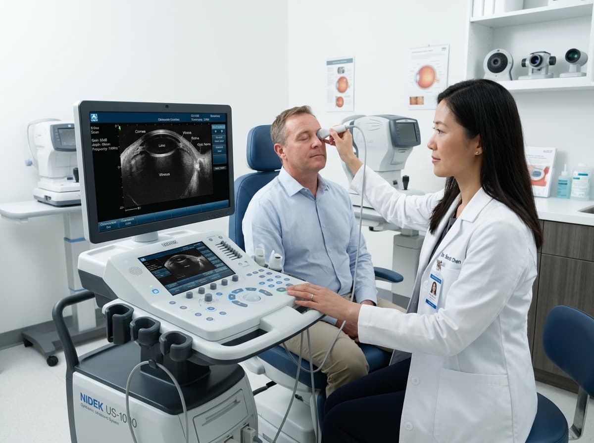
Uses high-frequency sound waves to visualize posterior ocular structures, confirming whether the retina is attached, identifying vitreous detachment, and ruling out intraocular tumors or trauma.
Patients when cloudy ocular media (dense cataract, corneal opacity, or vitreous hemorrhage) prevents direct ophthalmoscopic retinal inspection.
Why Choose The Eyes for this Procedure:
Common Symptoms & Clinical Indications
Patients suffering from these visual or physical complaints are typically recommended for this intervention:
Nidek / Quantel Medical High-Resolution 10MHz–20MHz B-Scan
Trans-palpebral high-frequency acoustic vector scan with cineloop playback
Step-by-Step Surgical & Diagnostic Protocol
01. Acoustic Probe Placement
A gentle ultrasound probe with acoustic coupling gel is placed over closed or open eyelids.
02. Multi-Vector Posterior Segment Sweeps
Transverse and longitudinal vector sweeps capture anterior and posterior vitreoretinal interfaces.
03. Real-Time Report Generation
High-resolution digital scan printout and consultant interpretation provided immediately.
Ready to Consult for B-Scan Ophthalmic Ultrasound?
Consultations are held at 162-A Shah Jamal Lahore. We allocate conflict-free 20-minute consultation slots with transparent, honest fees and zero corporate markups.